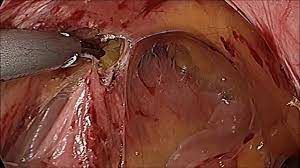
Bilateral Inguinal Hernia with Left sided Sliding Inguinal Hernia
Add to
Share
1,866 views
Report
2 years ago
Description
A sliding inguinal hernia is a protrusion of a retroperitoneal organ through an abdominal wall defect. The frequency of sliding hernias is estimated at 3-8% of all elective operations of inguinal hernias. Sliding hernias are supposed to be more anatomically challenging for a surgeon than uncomplicated non-sliding inguinal hernias. The anatomical and physiological concept of sliding inguinal hernia is frequently misunderstood by surgeons of all levels of experience. Not infrequently, any inguinal hernia that is big enough or has any organ (e.g. small intestine, sigmoid colon) inside its sac is referred to as a sliding hernia. Sliding hernias are known to surgeons for almost three centuries. The first description by Italian surgeon and anatomist from Pavia, Antonio Scarpa in 1809. they were feared as a complicated surgical condition. The main obstacle in the surgical approach to this type of hernia was-and still is-the fact that part of the hernia sac is, in reality, a retroperitoneal organ thus, during the opening of the sac inadvertent damage to a vital organ can be made. The advance of anatomical knowledge and the evolution of surgical techniques allowed for a better understanding of this entity. With a better understanding of the pathological anatomy of the sliding hernia various classification systems have been introduced. Currently, the best and most frequently used classification of sliding hernias is the one by Robert Bendavid. Bendavid divides the sliding inguinal hernia into three anatomical variants depending on the size of the sac and its relation to the sigmoid colon. Type I is defined as any hernia in which part of the peritoneal sac is made up by the wall of a viscus. Type II is defined as any hernia containing a retroperitoneal viscus and its mesentery, in which the mesentery forms part of the wall of the peritoneal sac. In type III the sliding hernia consists of a protrusion of a viscus itself, and the peritoneal sac is very small or even absent. This last variant is an extremely rare finding and accounts for approximately only 0.01% of all inguinal hernias. We strongly advocate the use of this classification in everyday practice as it enables surgeons to better understand the concept and hence better plan the operation of a sliding inguinal hernia. Schematic drawing of Bendavid Type I sliding inguinal hernia. The posterolateral aspect of the hernia sac is made up of the caecum and ascending colon. This type of sliding inguinal hernia accounts for almost 95% of all sliding inguinal hernia cases. The most common contents are sigmoid, caecum, and appendix. Type II sliding inguinal hernia. In this hernia, the mesentery forms part of the posterior wall of the sac, and part of the anterior wall of the caecum forms part of the posterior wall of the sac. This type of sliding inguinal hernia accounts for about 5% of all sliding inguinal hernia cases. The most common content is sigmoid. A sliding hernia is quite a common finding in infant girls: up to 20% of all hernias in this group of patients are sliding hernias containing ovary and fallopian tubes. In the adult population, almost all cases of sliding hernias are seen in men with only isolated reports of sliding inguinal hernias in women. The frequency of the sliding hernia in adults was historically estimated at around 6–8% of all hernia cases but more recent reports by our group estimate its frequency at 3.4%. Most probably it is due to the fact that today’s hernia patient present with smaller hernias with shorter duration of symptoms or even before the onset of symptoms. It is very rare to establish the preoperative diagnosis of a sliding inguinal hernia as there are no particular clinical signs indicating the possibility of a sliding hernia. Older patients with big hernias, presenting with a long history of inguinal lumps are the group most likely to have a retroperitoneal organ protruding into the hernia sac. In the literature, there are rare case reports of preoperative diagnosis of a sliding inguinal hernia containing urinary bladder based on a plain abdominal x-ray showing urinary bladder calculi within the groin. However, in the vast majority of cases, the diagnosis is made after the hernia sac is opened. If a surgeon does not open a hernia sac, a small sliding hernia can be easily overlooked. If the sac is manipulated gently this should not have any influence on the outcome of surgery in terms of early and late complications. As in the current practice, it is becoming increasingly rare for surgeons to routinely open an inguinal hernia sac, and a number of sliding hernias can undergo surgery without being recognized as such. Contact us World Laparoscopy Hospital Cyber City, Gurugram, NCR Delhi INDIA: +919811416838 World Laparoscopy Training Institute Bld.No: 27, DHCC, Dubai UAE: +971525857874 World Laparoscopy Training Institute 8320 Inv Dr, Tallahassee, Florida USA: +1 321 250 7653
Similar Videos


