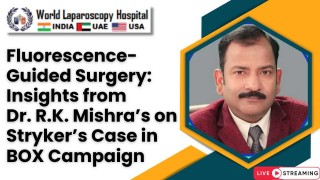
Indocyanine Green (ICG) in Bowel Perfusion: Dr. Deep Goyal’s Insights from WALS 2025
Add to
Share
114 views
Report
1 month ago
Description
The Role of Indocyanine Green (ICG) in Bowel Perfusion: Dr. Deep Goyal’s Insights from WALS 2025 At the World Association of Laparoscopic Surgeons (WALS) 2025 conference, held on February 2025, Dr. Deep Goyal, a renowned colorectal surgeon, shared groundbreaking insights into the transformative role of Indocyanine Green (ICG) in assessing bowel perfusion during laparoscopic surgery. His presentation highlighted how this innovative fluorescence imaging technique is revolutionizing intraoperative decision-making, particularly in colorectal procedures, and its potential to improve patient outcomes by reducing complications such as anastomotic leaks. Understanding ICG and Bowel Perfusion Indocyanine Green is a fluorescent dye that, when injected intravenously, binds to plasma proteins and emits a signal in the near-infrared spectrum when excited by a specialized light source. This property allows surgeons to visualize blood flow in real-time, offering a direct and objective assessment of tissue perfusion. In colorectal surgery, where the creation of a healthy anastomosis (the connection between two bowel segments) is critical, adequate blood supply is a key determinant of success. Poor perfusion can lead to anastomotic leakage, a devastating complication associated with increased morbidity, mortality, and prolonged hospital stays. Traditionally, surgeons have relied on subjective methods—such as observing bowel color, checking for bleeding at resection margins, or feeling for vessel pulsation—to assess perfusion. However, these approaches can be unreliable, especially in minimally invasive settings like laparoscopy, where tactile feedback is limited. Dr. Goyal emphasized that ICG provides a significant leap forward by offering a clear, real-time visualization of blood flow, enabling surgeons to make more informed decisions about where to resect and anastomose the bowel. Dr. Goyal’s Insights from WALS 2025 During his keynote address at WALS 2025, Dr. Goyal presented data from his extensive experience using ICG fluorescence angiography in laparoscopic colorectal resections. He underscored several key points: 1. Reducing Anastomotic Leaks: Dr. Goyal shared findings from his recent cases, where ICG was used to assess perfusion before and after anastomosis. In instances where fluorescence revealed suboptimal blood flow, he adjusted the resection line to better-perfused tissue, potentially averting leaks. He noted that while large-scale randomized trials are still needed, his observations align with emerging evidence suggesting that ICG can lower leak rates, particularly in high-risk procedures like low anterior resections. 2. Real-Time Decision-Making: One of the standout advantages of ICG, according to Dr. Goyal, is its immediacy. After injecting a small bolus of ICG (typically 0.2-0.4 mg/kg), fluorescence becomes visible within seconds using modern laparoscopic systems, such as those equipped with near-infrared cameras. This allows surgeons to assess perfusion intraoperatively and adapt their approach on the spot—whether by resecting additional tissue or opting for a protective stoma in cases of questionable viability. 3. Integration with Robotic Surgery: Dr. Goyal highlighted the synergy between ICG and robotic platforms like the da Vinci system, which many laparoscopic surgeons now use. These systems feature integrated fluorescence imaging, allowing seamless transitions between standard and near-infrared views. He showcased footage from a robotic low anterior resection where ICG clearly delineated a perfusion deficit, guiding a critical adjustment that he believes prevented a postoperative complication. 4. Challenges and Future Directions: While enthusiastic about ICG’s potential, Dr. Goyal was candid about its limitations. He noted that interpreting fluorescence remains somewhat subjective without standardized quantitative tools. To address this, he advocated for the development of software to measure fluorescence intensity and perfusion patterns objectively—a step he believes could further enhance ICG’s reliability. Additionally, he called for more robust, multicenter studies to solidify its role in routine practice and justify its cost. Practical Applications in Laparoscopic Surgery Dr. Goyal illustrated ICG’s utility with a case study from his practice: a 62-year-old patient undergoing a laparoscopic sigmoidectomy for cancer. After dividing the mesentery, he injected ICG and observed fluorescence using a near-infrared laparoscope. The imaging revealed a segment of the proximal bowel with delayed fluorescence, indicating poor perfusion. Based on this, he resected an additional 5 cm of colon to reach well-perfused tissue, then confirmed adequate blood flow at the anastomosis site post-construction. The patient recovered without complications, reinforcing Dr. Goyal’s confidence in the technique. He also touched on ICG’s broader applications beyond perfusion, such as lymphatic mapping and ureter visualization, but emphasized that its most immediate impact lies in ensuring anastomotic integrity. “For colorectal surgeons, the anastomosis is the moment of truth,” he said. “ICG gives us a window into that truth like never before.”
Similar Videos