Giant Gallbladder: Surgical Challenges and Transition to Open Surgery
Add to
Share
102 views
Report
1 month ago
Description
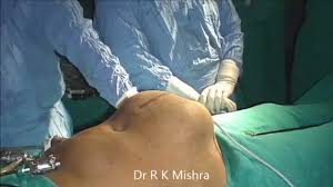
Introduction The gallbladder, a small pear-shaped organ nestled beneath the liver, plays a vital role in digestion by storing and concentrating bile. While gallbladder diseases such as cholelithiasis (gallstones) and cholecystitis (inflammation) are relatively common, encountering a "giant gallbladder" — an abnormally enlarged gallbladder significantly exceeding its typical size — is exceedingly rare. Such cases present unique diagnostic and surgical challenges, often requiring tailored approaches due to anatomical distortions, obscured landmarks, and heightened risks of complications. This case report, presented as part of the World Association of Laparoscopic Surgeons (WALS) 2025 conference, explores the complexities of managing a giant gallbladder, focusing on the surgical challenges encountered during a laparoscopic procedure and the critical decision to transition to open surgery. Dated March 01, 2025, this report underscores the evolving landscape of minimally invasive surgery while highlighting the importance of adaptability in the operating theater. Case Presentation The patient, a 62-year-old female, presented to the emergency department with a three-day history of worsening right upper quadrant pain, nausea, and low-grade fever. Her medical history included hypertension and obesity (BMI 34), but no prior abdominal surgeries or known gallbladder disease. Initial laboratory findings revealed mild leukocytosis (WBC 13,000/µL) and elevated liver enzymes (ALT 85 U/L, AST 70 U/L), suggestive of an inflammatory process. An abdominal ultrasound was performed, revealing a markedly distended gallbladder measuring approximately 28.0 x 16.0 cm, filled with sludge and multiple gallstones, but no clear evidence of perforation or pericholecystic fluid. The initial radiologic impression was a "giant celiac cyst" due to the organ’s enormous size and cystic appearance in the hepatic region. Given the suspicion of acute cholecystitis, the surgical team planned a laparoscopic cholecystectomy, the gold standard for gallbladder removal. The patient was counseled on the procedure, including the possibility of conversion to open surgery if necessitated by intraoperative findings. After obtaining informed consent, she was taken to the operating room under general anesthesia. Surgical Approach and Challenges Laparoscopic cholecystectomy was initiated with standard port placement: a 10-mm umbilical port for the laparoscope and three additional 5-mm ports in the epigastric and right subcostal regions. Upon insufflation with carbon dioxide, the abdominal cavity was visualized, revealing a grossly enlarged gallbladder occupying much of the right upper quadrant and extending inferiorly toward the pelvis. The organ’s size distorted nearby anatomical landmarks, including the cystic duct, cystic artery, and hepatocystic triangle (Calot’s triangle), critical structures that must be clearly identified to safely perform the procedure. Several challenges emerged during the laparoscopic approach: 1. Visualization Difficulties: The giant gallbladder obscured the operative field, making it nearly impossible to achieve the "critical view of safety" — a technique requiring clear visualization of the cystic duct, cystic artery, and gallbladder detachment from the liver bed before clipping or transecting any structures. The sheer volume of the organ compressed adjacent structures, complicating dissection. 2. Limited Instrument Reach: Standard laparoscopic instruments struggled to maneuver around the massive gallbladder. Retraction was inadequate, as the organ’s weight and size resisted manipulation with graspers, increasing the risk of inadvertent perforation or bile spillage. 3. Risk of Vascular Injury: The cystic artery, typically a small vessel, was difficult to isolate due to its displacement by the enlarged gallbladder. Prolonged dissection in this confined space raised concerns about potential injury to the hepatic artery or portal vein, both of which lie in close proximity. 4. Gallbladder Wall Integrity: The gallbladder wall appeared thinned and friable, likely due to chronic inflammation and distension. This heightened the risk of rupture during manipulation, which could lead to bile peritonitis or dissemination of gallstones into the peritoneal cavity. 5. Time Constraints: The prolonged operative time required to address these challenges increased the patient’s exposure to anesthesia and the risk of complications such as pneumoperitoneum-related issues (e.g., hypercapnia or gas embolism). Despite meticulous efforts over 90 minutes, the surgical team could not safely proceed laparoscopically. The decision was made to convert to an open cholecystectomy to ensure patient safety and complete the procedure effectively. Transition to Open Surgery The laparoscopic ports were removed, and a right subcostal incision (approximately 15 cm) was made to access the abdominal cavity directly. Upon opening the abdomen, the giant gallbladder was immediately apparent, confirming the ultrasound findings. The organ was carefully mobilized, revealing a patent cystic duct with no obstructing stones or masses, suggesting a congenital etiology for its size rather than an acquired obstruction. The gallbladder was aspirated to remove approximately 1.2 liters of bile and sludge, reducing its volume and facilitating dissection. With improved visualization and tactile feedback, the surgical team successfully identified and ligated the cystic duct and artery. The gallbladder was then dissected from the liver bed using a combination of sharp dissection and electrocautery. No significant bleeding or bile leakage occurred, and the procedure was completed without further complications. The specimen was sent for histopathological analysis, which later confirmed chronic cholecystitis with cholelithiasis, and no evidence of malignancy. Postoperative Course The patient was monitored in the surgical ward for five days. She experienced moderate postoperative pain, managed with intravenous analgesics, and developed a transient ileus that resolved by postoperative day 3. Drains placed during surgery showed minimal output and were removed prior to discharge. Antibiotics were continued for 48 hours to prevent infection. At a two-week follow-up, the incision was healing well, and the patient reported resolution of her preoperative symptoms. Conclusion The surgical management of a giant gallbladder exemplifies the delicate balance between the benefits of minimally invasive techniques and the realities of complex anatomy. In this WALS 2025 case report, the transition from laparoscopic to open cholecystectomy was a critical step to overcome visualization difficulties, technical limitations, and heightened risks. As laparoscopic surgery continues to evolve, lessons from such cases will inform future innovations, ensuring that surgeons are equipped to handle even the most atypical presentations. For now, the ability to adapt remains a cornerstone of surgical excellence, particularly when faced with the rare and formidable challenge of a giant gallbladder.
Similar Videos


