Laparoscopic Nephrectomy Lecture by Dr R K Mishra
Add to
Share
3,676 views
Report
Description
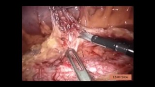
This video is of Laparoscopic Nephrectomy Lecture by Dr R K Mishra. A laparoscopic nephrectomy is minimally invasive operation to remove kidney. A laparoscopic nephrectomy involves removing an entire kidney through keyhole incisions in the flank, the side of the body between the ribs and the hip. A laparoscopic nephrectomy removes the kidney by using laparoscopic equipment. Long thin instruments are passed through up to five small incisions made in the flank, each about 1cm in length. The abdomen is first filled with carbon dioxide, which separates the tissues to allow for vision during the surgery. A camera is then passed, giving the urologist a detailed picture inside the abdomen. The other incisions are used to pass cutting and suturing instruments so the blood supply to the kidney can be isolated and tied off and the kidney removed either with or without its surrounding structures. A wound drain is then inserted to drain any wound ooze. This is usually stitched in place and stays in for 1 – 2 days. A nephrectomy is usually done for one of two reasons, either for cancer of the kidney or because of a non- functioning kidney. In the case of kidney cancer a radical laparoscopic nephrectomy is done. This is done in an attempt to rid the body of cancer by removing the entire kidney and adrenal gland, with its surrounding fat and attached vessels. In more advanced cases it may be done to stop continued bleeding from the effected kidney. For non-functioning kidneys, which are either caused by large stones, a lack of blood supply or abnormal kidney structure, a simple laparoscopic nephrectomy is done. This is where only the kidney itself is taken and the adrenal gland and other structures are left behind. A simple nephrectomy is usually done to avoid recurrent infection and pain and the possibility of severe illness because of infection.
Similar Videos